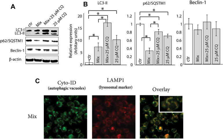
Fig. 2. Effect of co-administration of C6-ceramide and tamoxifen on induction of autophagic flux in KG-1 cells.
A) Impact of treatment on expression of LC3-II, p62, and Beclin-1. Cells were treated with C6-ceramide-tamoxifen for 18 h and then assessed for LC3-II, p62, and Beclin-1 expression by Western blot. B) Densitometric quantitation using ratios of LC3-II, p62, Beclin-1, to β-actin. C) Effect of treatment regimen on fusion of lysosome with autophagosome. MEF cells transfected with lysosomal marker LAMP1-RFP (red fluorescent protein) were used. After treatment (18 h), MEF LAMP1+/+ cells were stained with Cyo-ID to identify autophagosomes. (For interpretation of the references to color in this figure legend, the reader is referred to the Web version of this article.)

