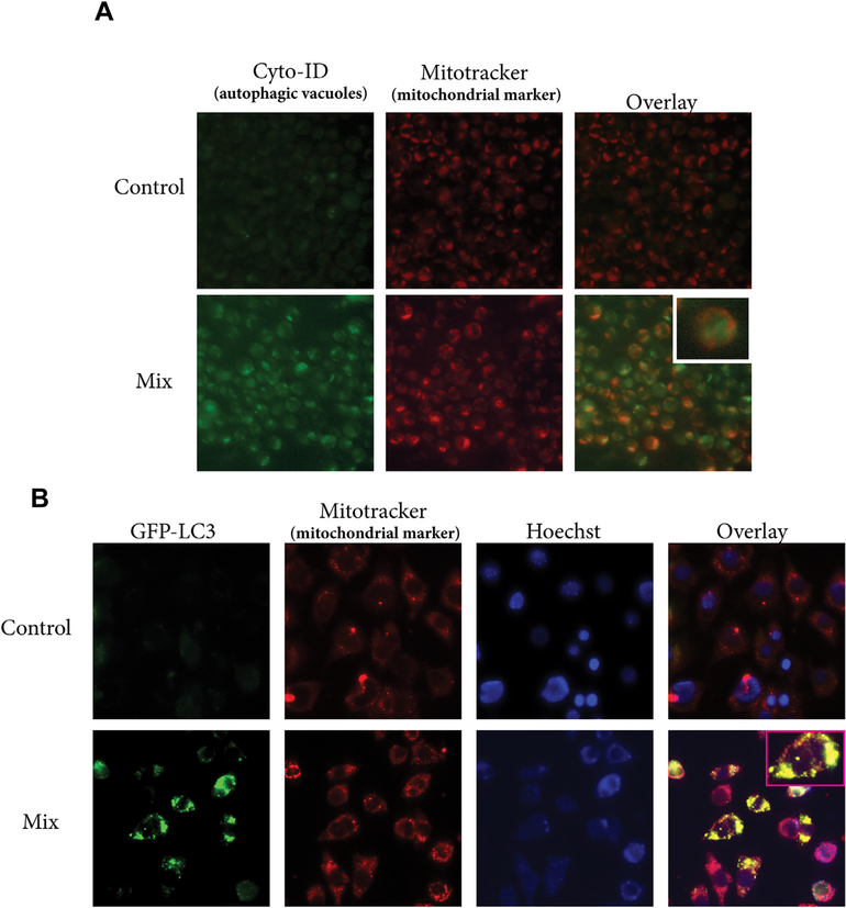
Fig. 4. C6-ceramide-tamoxifen combination induces mitophagy.
A) Effect of drug mix on co-localization of autophagosome and mitochondria. KG-1 cells were exposed to DMSO (control) or to the drug mix (18 h), after which, cells were stained with Cyto-ID and Mitotracker Red. The fluorescence photomicroscopy overlay denotes co-incidental staining between autophagosome and mitochondria in cells exposed to mix (C6-ceramide-tamoxifen). B) GFP-LC3-MEF cells were exposed to vehicle control (DMSO) or the drug mix for 18 h, followed by staining with Mitotracker Red and Hoechst (nucleus staining). Images were obtained by fluorescence photomicroscopy. The mix overlay denotes co-localization of GFP-LC3 with mitochondria. (For interpretation of the references to color in this figure legend, the reader is referred to the Web version of this article.)

