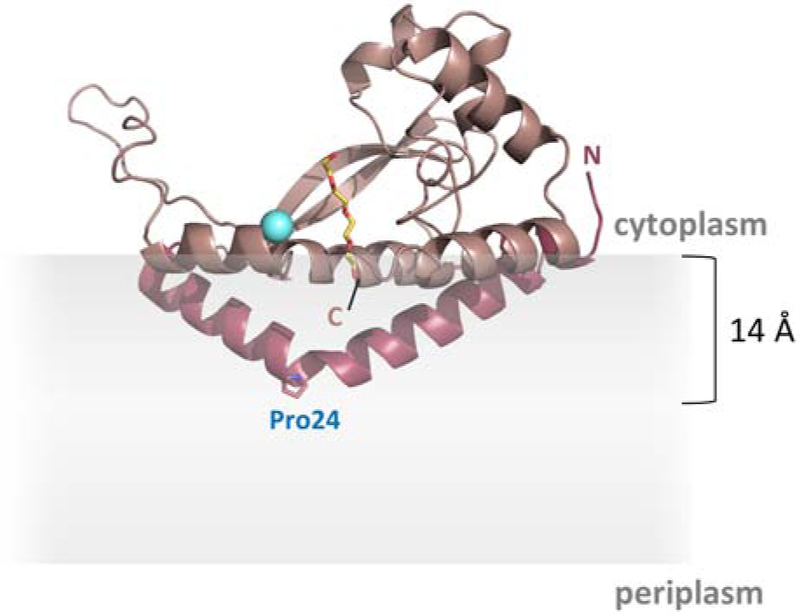
Fig. 2 – Structure of PglC.

The reported structure of PglC (PDB 5W7L) [35]. The N-terminal reentrant helical domain is shown in dark pink. Pro24 in the membrane-resident domain is shown as sticks. The Mg2+ in the active site is shown in cyan. A co-crystallized PEG molecule marking the putative PrenP-binding site is shown in yellow. The location of the membrane, as calculated in [35], is represented in gray.
