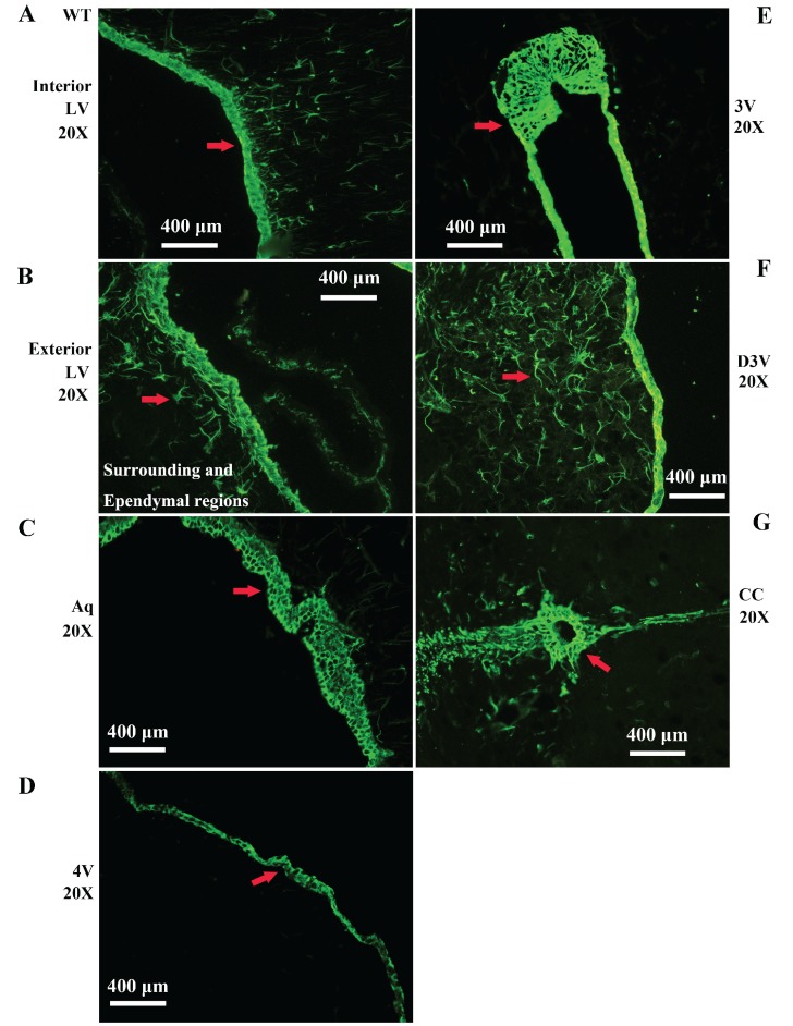
Figure 2.
Representative images of VPCs distribution in different ventricular systems in adult WT mice. A, B The VPCs distribution in LV. The VPCs in LV mainly distributed in the interior (A), the exterior and the surrounding and Ependymal regions (B). C The VPCs distribution in Aq. VPCs in Aq mainly distributed in the ependymal region. D The VPCs distribution in 4V. VPCs in 4V mainly distributed in the ependymal region. E The VPCs distribution in 3V. VPCs in 3V mainly distributed in the ependymal region. F The VPCs distribution in D3V. VPCs in D3V mainly distributed in the ependymal region. G The VPCs distribution in CC. VPCs in CC mainly distributed in the ependymal region. The red arrow marked the positive cell, bar = 400µm.

