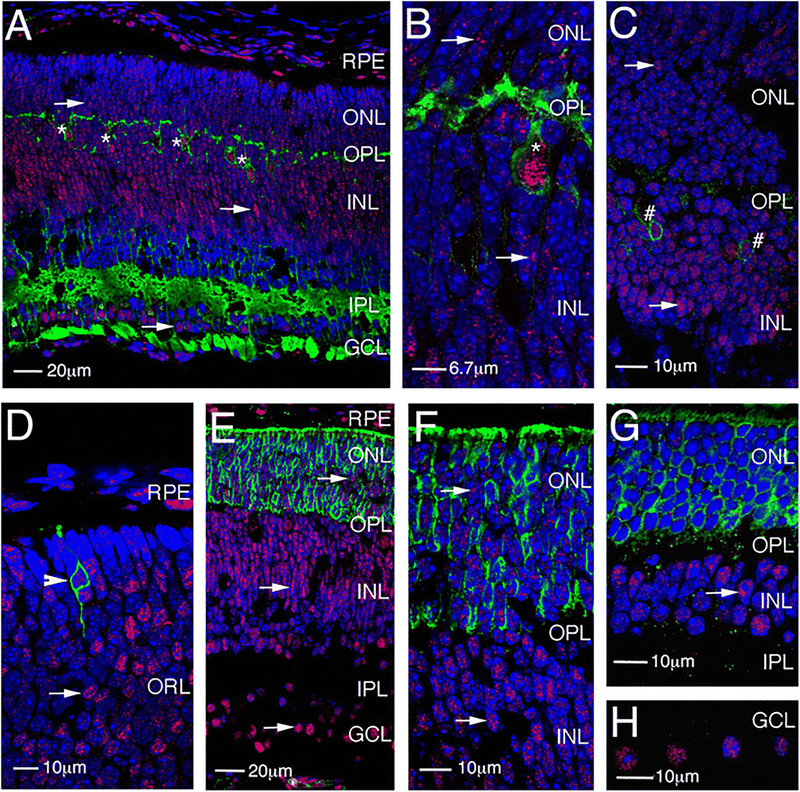
Fig. 2.
LSD1 expression in different retina cell types. a, b Immunofluorescence microscopic images of sections of mouse retina at PN7 stained with HNK-1 (green), LSD1 (red), and nuclear counterstained with Hoechst33358 (blue). c Immunofluorescence microscopic images of sections of mouse retina at PN7 stained with PKCα (green), LSD1 (red), and nuclear counterstained with Hoechst33358 (blue). d–h Immunofluorescence microscopic images of sections of mouse retina stained with Rhodopsin (green), LSD1 (red), and nuclear counterstained with Hoechst33358 (blue) at PN1 (d), PN7 (e, f), and PN28 (g, h). RPE, retinal pigment epithelium; GCL, ganglion cell layer; OPL, outer plexiform layer; IPL, inner plexiform layer; ONL, outer nuclear layer; INL, inner nuclear layer. Arrows show LSD1 expression in different retinal layers

