Fig. 4.
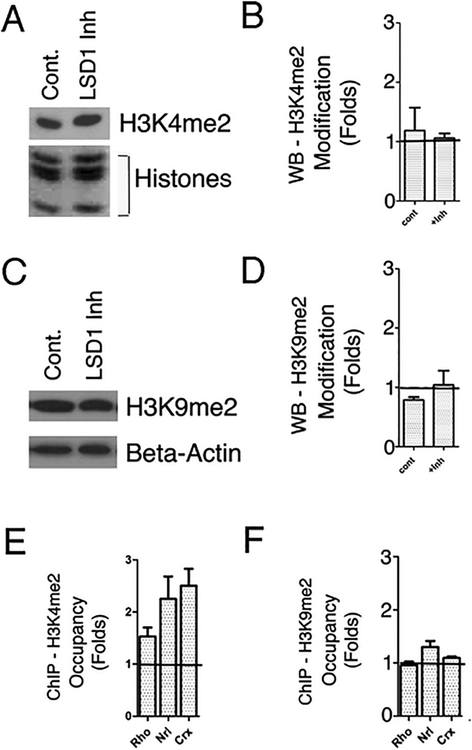
Global level of LSD1 substrates, H3K4me2 and H3K9me2 during LSD1 inhibition. a Western blot of samples isolated from mouse PN1 retinal explants cultured for 8 days with or without LSD1 inhibitor probed with antibody against H3K4me2. Coomassie staining of core histones was used as loading control. b Quantification of H3K4me2 Westerns, n=3. c Western blot of samples isolated from mouse PN1 retinal explants cultured for 8 days with ±LSD1 inhibitor probed with antibody against H3K9me2. Beta-actin was used as loading control. d Quantification of H3K9me2 Westerns, n=3. e, f. Comparison of H3K4me2 (e) or H3K9me2 (f) accumulation on gene promoters in 8 days retina explants cultured with TCP vs. with control media only. ChIP experiments were done on 2–3 biological replicas and quantified by real-time PCRs, y-axis represent fold changes relative to control

