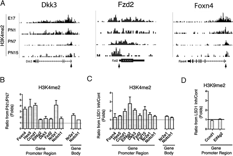
Fig. 6.
Levels of H3K4me2 maintained over progenitor genes under LSD1 inhibition. a Combined genome-wide tracks of H3K4me2 accumulation at different stages of normal retina development for factors important for general retinogenesis and cell type specification; gene TSS is marked by arrow. b Comparison of H3K4me2 accumulation on promoter and body at PN1 and PN7 during normal retina development for selected genes upregulated after LSD1 inhibition. ChIP experiments were done in 2–3 biological replicates; quantitative real-time PCRs were performed with primers specific for promoter and gene body (Online Resource Table 1) of progenitor genes. c–d Comparison between H3K4me2 (c) and H3K9me2 (d) accumulation on gene promoters and gene body areas in retina explants cultured for 8 days with TCP or in media only. ChIP experiments were done in 2–3 biological replicates; quantitative real-time PCRs were done with same primers as in (b)

