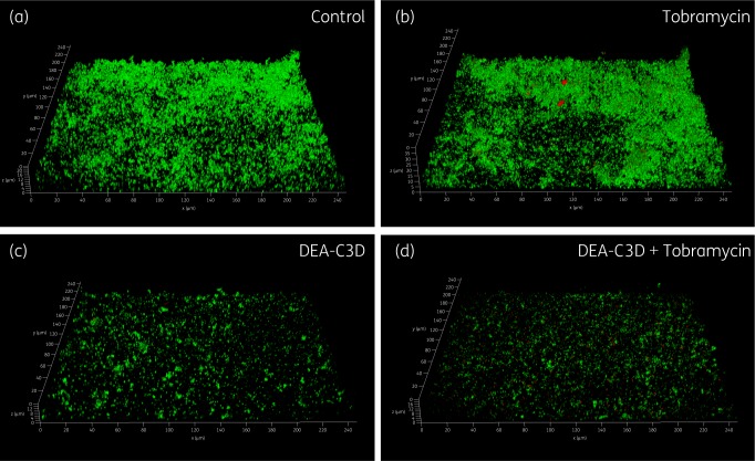
Figure 4.
Appearance of P. aeruginosa biofilms following tobramycin and DEA-C3D treatments, alone and in combination. Biofilms formed by P. aeruginosa clinical CF isolate PA68 were treated with 4 mg/L tobramycin (b), 256 μM DEA-C3D potassium salt (c) or DEA-C3D/tobramycin combination (d), and compared with untreated control biofilms (a). Biofilms were stained with SYTO9 (green=viable cells) and propidium iodide (red=dead/damaged cells) before CLSM. Representative 3D images of each treatment are shown; x- and y-axes=246 μm by 246 μm. Experiments were carried out in duplicate and showed similar results.

