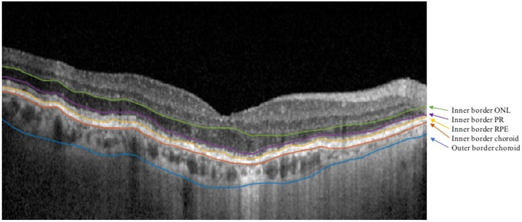
Figure 1: Retinal Layer Segmentations.
SD-OCT B-scan image in an eye of a patient with GA. The B-scan shows manually segmented and smoothed borders separating the layers of interest (arrows). For simplicity, the outer border of the outer nuclear layer (ONL) is the inner border of the photoreceptor (PR) layer (purple); the outer border of the PR layer is the inner border of the retinal pigment epithelium (RPE) (yellow), and the outer border of the RPE is the inner border of the choroid (red).

