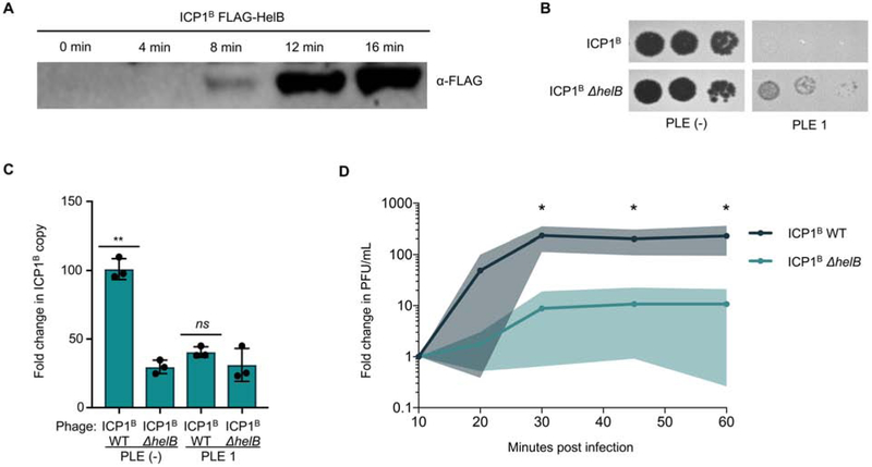
Figure 5. Loss of helB permits escape from PLE but leads to a defect in ICP1 fitness.
A, Western blot of endogenously FLAG-tagged HelB following infection of PLE (−) V. cholerae. B, Tenfold ICP1 dilutions spotted on the listed V. cholerae lawns. C, Fold change in ICP1 copy number following infection of the listed V. cholerae host as measured by qPCR. D, Relative burst size of ICP1B and ICP1B ΔhelB on PLE (−) V. cholerae as measured by one-step growth curves. *p<0.05, **p<0.001, ns not significant.

