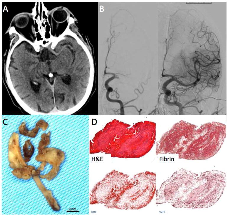
FIGURE 1.
Clot Attenuation and Composition and Revascularization: Fibrin Rich Clot. 67-year-old female with acute onset right sided hemiparesis and aphasia.
A. Non-contrast CT using thin-section 1mm reconstruction shows no hyperdense vessel.
B. Left ICA cerebral angiogram shows occlusion of the left MCA. Four passes with a Solitaire 4×40 were needed to open the vessel.
C. Gross photo of the retrieved clot shows white clot consistent with fibrin rich thrombus.
D. H&E stain shows that a majority of the clot was composed of fibrin, the overall clot composition was 90% fibrin and 7%RBCs.

