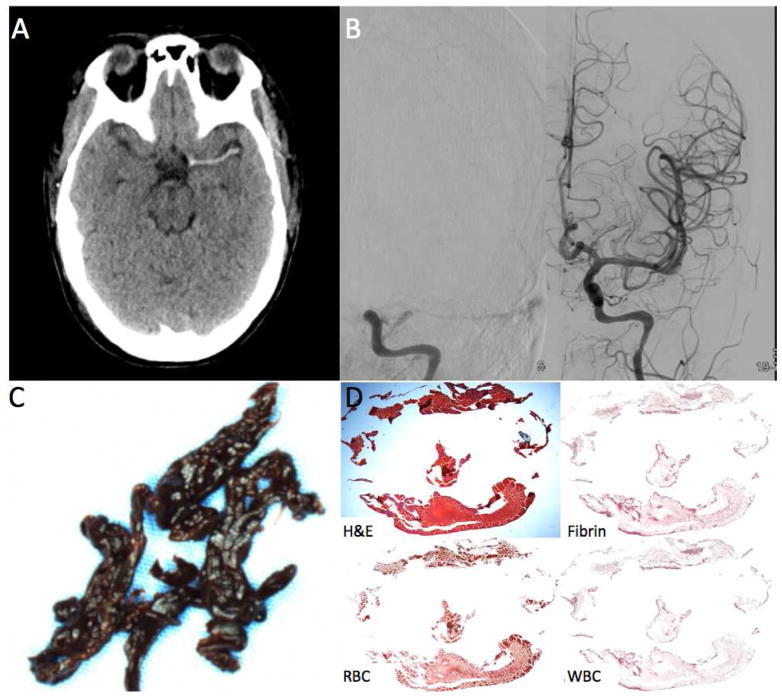
FIGURE 4.
Clot Attenuation and Composition and Revascularization: RBC Rich Clot. A 24 year old female status post aortic root replacement who was not waking up two hours after surgery.
A. Non-contrast CT shows a long hyperdense thrombus of the left MCA.
B. The patient was taken straight to angiography which confirmed the left MCA occlusion. One pass with a 6cm Solitaire was needed to revascularized the occlusion.
C. Gross photo of the clot shows dark red clot consisted with an RBC rich thrombus.
D. H&E stain shows that a majority of the clot was composed of RBCs (94% RBC density).

