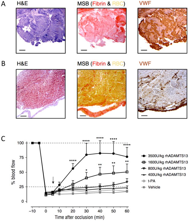
FIGURE 7.
VWF in ischemic stroke thrombi.
Intracranial thrombi retrieved from stroke patients that underwent thrombectomy procedure were collected for histological analysis. Consecutive thrombi sections were stained with H&E, Martius Scarlet Blue (MSB) and anti-VWF antibodies. Classical H&E staining show overall thrombus composition and organization. On MSB staining, red areas show the presence of fibrin whereas red blood cells appear yellow. Varying amounts of VWF (brown color) were found in all the thrombi Two representative patient thrombi are shown illustrating a VWF-rich thrombus (A) and a RBC-rich, VWF-poor thrombus (B). Scale bar: 50μm. (C) An occlusive thrombus was generated in the right MCA of C57Bl/6J mice. Five minutes after occlusion, vehicle, t-PA or different doses of rhADAMTS13 were intravenously administered (arrow) and MCA blood flow was monitored for 60 minutes. Average blood flow profiles show that rhADAMTS13 restores MCA blood flow in a dose dependent way, while t-PA was unable to restore blood vessel patency. (n = 10 and 8 mice respectively for vehicle and 3500U/kg rhADAMTS13, n = 5 for the lower doses of rhADAMTS13 and t-PA; *, p < 0.05; **, p < 0.01; ****, p < 0.001; compared with vehicle).
Adapted from Denorme F, Langhauser F, Desender L, et al. ADAMTS13-mediated thrombolysis of t-PA-resistant occlusions in ischemic stroke in mice. Blood. 2016;127(19):2337–2345; with permission.

