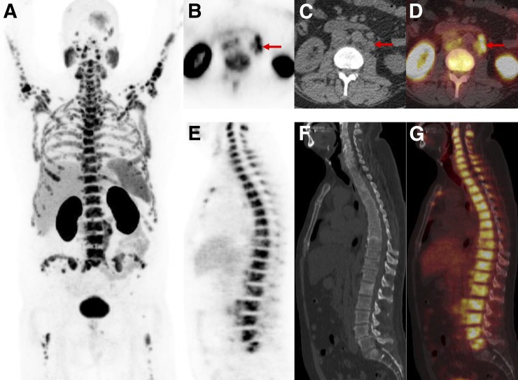
FIGURE 10.
PSMA-RADS-5: images of patient with extensive metastatic PCa. (A) Whole-body maximum-intensity-projection image shows diffuse osseous metastatic disease and retroperitoneal adenopathy. This scan would be categorized as PSMA-RADS-5, and there are also several individual PSMA-RADS-5 lesions. (B–D) Axial CT (B), axial 18F-DCFPyL PET (C), and axial 18F-DCFPyL PET/CT (D) images through retroperitoneum show multiple enlarged lymph nodes (short-axis diameter, >1.0 cm) with intense uptake (arrows). (E–G) Sagittal bone window CT (E), sagittal 18F-DCFPyL PET (F), and sagittal 18F-DCFPyL PET/CT (G) images show diffuse metastatic disease in spine with intense uptake and underlying sclerotic changes in bones. Lack of sacral uptake in F and G is due to previous pelvic radiation therapy.

