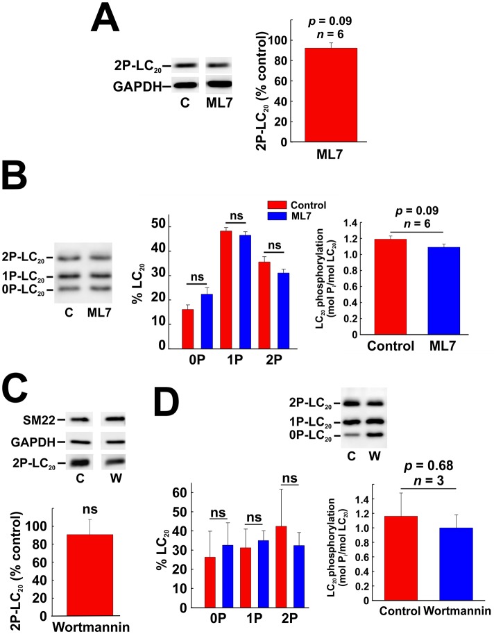
Fig 8. Effect of inhibition of MLCK on phosphorylation of LC20 in coronary arterial smooth muscle cells.
(A) Effect of ML7 (10 μM) pre-treatment on serum-induced LC20 diphosphorylation (analysed by western blotting with anti-2P-LC20; left-hand panel) with quantitative analysis (right-hand panel). Statistical analysis was carried out with Student’s unpaired t-test. (B) Effect of ML7 (10 μM) pre-treatment on LC20 phosphorylation (analysed by Phos-tag SDS-PAGE and western blotting with anti-LC20) following 2 min of serum stimulation. Controls (C) were incubated with vehicle rather than ML7. A representative western blot is shown in the left-hand panel, quantification of unphosphorylated, mono- and diphosphorylated LC20 in the middle panel and overall phosphorylation stoichiometry in the right-hand panel (n = 6). Statistical analysis was carried out with Student’s unpaired t-test. (C) Effect of wortmannin (1 μM) pre-treatment on serum-induced LC20 diphosphorylation (analysed by western blotting with anti-2P-LC20; upper panel) with quantitative analysis (n = 3; lower panel). (D) Effect of wortmannin (1 μM) pre-treatment on serum-induced LC20 diphosphorylation (analysed by Phos-tag SDS-PAGE with anti-LC20; upper panel) with quantitative analysis (lower panels). “ns” denotes p > 0.05 (Student’s unpaired t-test).

