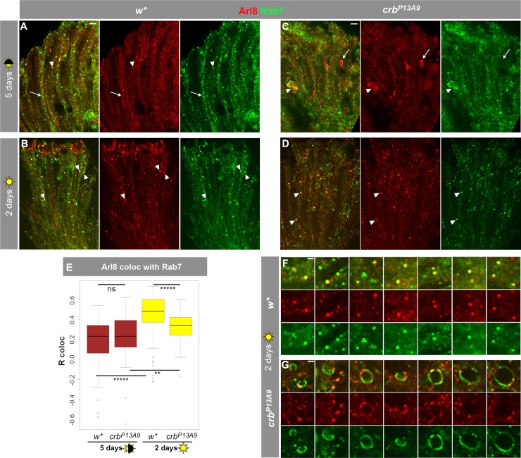
Fig 5. Exposure to constant light causes increased co-localization of Arl8 with late endolysosomal Rab7 and abnormal Rab7 compartment shapes in crbP13A9.
(A, C) w* and crbP13A9 retinas, respectively, under 5 days normal light conditions (12h light, 12h dark) show some overlap of large endogenous Rab7-carrying vesicles (green) with Arl8 (red) (arrowhead). Additionally, many normal-sized compartments in w* (A) and abnormal patches in crbP13A9 (C) are non-colocalizing (arrows), resulting in a relatively low, positive Pearson correlation coefficient (R coloc) of ~0.25 (on a scale of -1 to +1) for both genotypes over the whole retina (E, brown boxes). (B, D) Constant light stress causes an increase in co-localization between Rab7 and Arl8 (arrowheads) in w* and crbP13A9 retinas, respectively (R coloc = ~0.5 for w*; ~0.33 for crbP13A9; E). Scale bars for A-D: 5 μm. (E) Quantification of Rab7 and Arl8 co-localization. 5 days of normal light conditions does not reveal any significant difference in Arl8/Rab7 co-localization (brown box plots). Two days of constant light stress causes a significant increase in the Pearson correlation coefficient in w*, and a smaller but significant increase in crbP13A9. Quantification was done on ROIs from image stacks, as described in Fig 2. For 5 days normal light, w* n = 110 images and crbP13A9 n = 118 images; for 2 days constant light, w* n = 108 images and crbP13A9 n = 105 images. (F, G) Enlarged images showing numerous large round lysosomal compartments in w* positive for both Arl8 and Rab7. In contrast, large, distended Rab7-positive compartments in crbP13A9 can be seen in contact with small foci of Arl8 (G). Scale bars: 1 μm.

