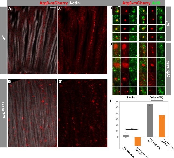
Fig 6. Autophago-lysosomal marker Atg8-mCherry loses its association with Arl8 in crbP13A9 mutant retinas.
(A-B’) Longitudinal optical sections of w* (A, A’) and crbP13A9 (B, B’) retinas of flies expressing Rh1-Gal4-mediated Atg8-mCherry (red). Retinas of flies kept for 6 days at 12h light/12h dark show differences in morphology of autophago-lysosomes in crbP13A9 (B, B’). Rhabdomeres are labelled for F-actin (phalloidin; white). Scale bar: 5 μm. (C, D) Examples of single autophagosomal Atg8-mCherry compartments (red) in w* (C) and crbP13A9 (D). Atg8-mCherry is often directly adjacent to or surrounded by Arl8 (green) in the control (C), whereas it is non-overlapping with Arl8 in crbP13A9 (D). Scale bars: 1 μm. (E) Quantification showing loss of Atg8-mCherry (driven by Rh1-Gal4) co-localization with Arl8 in crbP13A9 in individual compartments, similar to those shown in C and D) measured by Pearson’s R coloc and Manders’ thresholded co-localization coefficient tM2 for the Atg8-mCherry channel. Total compartment number (n) for w* = 26, n for crb P13A9 = 62.

