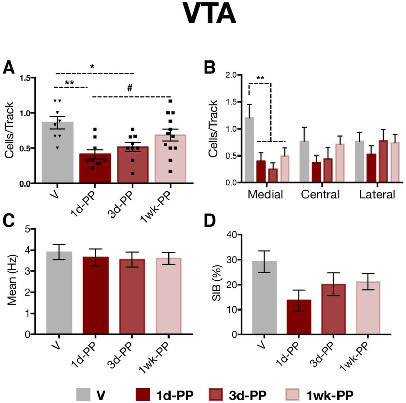
Figure 4. Reduced VTA DA population activity during the first 3 days postpartum.
A) 1- and 3-day postpartum females exhibited a reduction in the number of spontaneously active DA cells in the VTA (i.e. population activity) compared with virgins (p < 0.05). B) Early postpartum females exhibited a selective attenuation of DA neuron activity in the medial aspect of the VTA (p <0.01). C,D) No changes between virgins and early postpartum females were observed in C) firing rate (p=0.95) or D) percentage of spikes firing in bursts (p=0.08). *p < 0.05, ** p < 0.01, #p=0.06, Error bars represent mean ± SEM. Gray bars represent virgins (n=8) and red bars represent postpartum females (n=9-12 per group).

