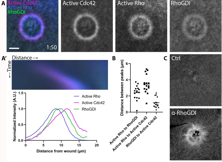
Figure 4. RhoGDI is recruited to single-cell wounds enriched in Rho and Cdc42 activity.
(A) Oocytes microinjected with wGBD (magenta), rGBD (blue) and GDI (green); (A’) Kymograph of wound closure and line scan of radially-averaged fluorescence intensity from (A); (B) Quantification of distance between peaks (n = 16); (C) Wounded oocytes fixed and stained with anti-X. laevis GDI. Scale bar 10 μm, time min:sec.
Figure 4—figure supplement 1. X. laevis RhoGDI antibody specificity.


