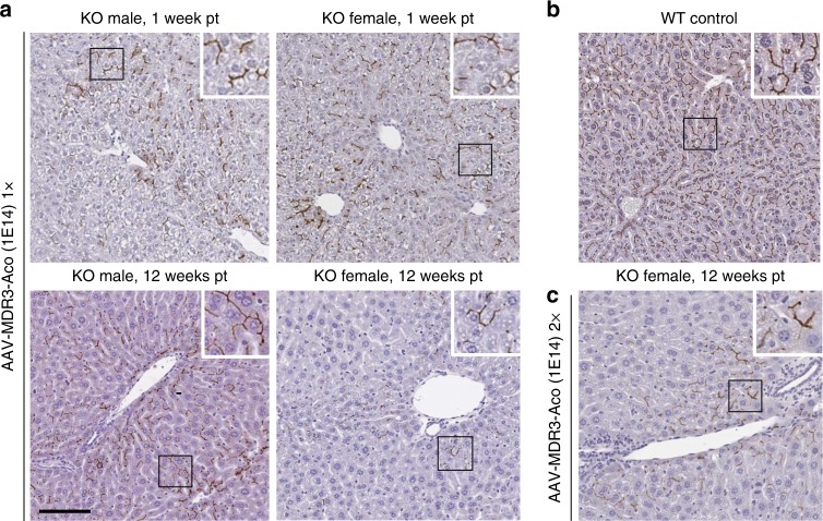
Fig. 2. Analysis of AAV-MDR3-Aco expression in Abcb4−/− mice.
Two-week-old Abcb4−/− mice (KO) mice received one (a) or two (c) doses of 1 × 10¹⁴ VG/kg of AAV-MDR3-Aco, and MDR3 expression was analysed at the indicated times after the first dose (pt) by IHC with an anti-MDR3-specific antibody. In c, the second dose was given when mice were 5 weeks old. A female WT mouse is shown as positive control for MDR2 staining (b). Representative pictures from one mouse in each group are shown. Scale bar = 100 μm.

