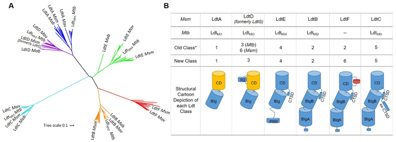
Figure 1.
Phylogenetic analysis and reclassification of mycobacterial Ldts based on structure, sequence, and phylogeny. A) Mycobacterial Ldts cluster into six clades that confirm five previously identified Ldt classes: class 1 (blue), class 2 (orange), class 3 (purple), class 4 (green) and class 5 (cyan). Msm LdtF clusters into a distinctive sixth clade (red). Mav = Mycobacterium avium, Mab = Mycobacterium abscessus. B) Reclassification and cartoon depiction of mycobacterial Ldts. CD = catalytic domain, BIg = bacterial Ig-like domain, R2 = Region 2 sequence (Figures S2 and S3), PRR = proline-rich region, CTSD = C-terminal subdomain, ex-CTSD = extension of the CTSD. Yellow CDs indicate the presence of Region 1 sequence (Figure S3). Class 6 Ldts contain a 10-residue insertion (red cylinder in CD). Classes 2, 5, and 6 contain an N-terminal lipobox.19 Asterisk refers to reference 19. Classes are arranged to indicate increasing apparent structural complexity.

