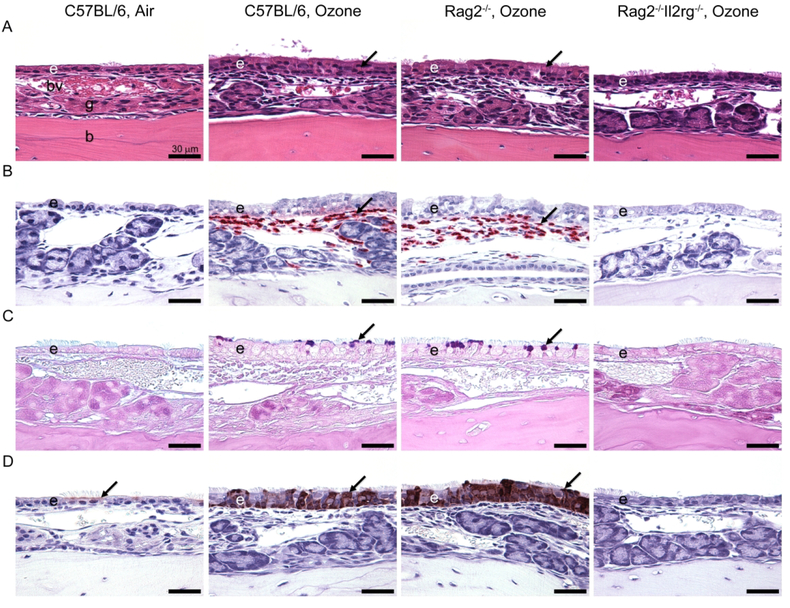
Figure 2.
Light photomicrographs of eosinophilic inflammation and epithelial remodeling in the nasal mucosa lining the lateral wall of the proximal nasal passages after repeated ozone exposure. (A) Hematoxylin and eosin stain. Exposure of 0.8 ppm ozone for 9 days caused marked rhinitis and epithelial remodeling (i.e., hyperplasia, mucous cell metaplasia, and hyalinosis) in innate lymphoid cell-sufficient C57BL/6 mice and Rag2−/− mice. By contrast, innate lymphoid cell-deficient Rag2−/−Il2rg−/− mice exposed to ozone had no nasal lesions. Arrows indicate eosinophilic globules (hyalinosis) in the transitional epithelium. (B) Immunohistochemical stain for major basic protein in murine eosinophils. Large numbers of eosinophils (arrows) were detected in the nasal mucosa of C57BL/6 mice and Rag2−/− mice after 9-day exposure to ozone. (C) Alcian blue (pH 2.5)/periodic acid–Schiff stain for acidic and neutral mucosubstances. C57BL/6 and Rag2−/− mice exposed to ozone for 9 days stored Alcian blue (pH 2.5)/periodic acid–Schiff–stained intraepithelial mucosubstances in the apical aspect of nonciliated epithelial cells (arrows). (D) Immunohistochemical stain for chitinase-like 3 (YM1) and chitinase-like 4 (YM2) proteins. Air-exposed mice contained small amounts of YM1/YM2 proteins in the apical aspect of ciliated epithelial cells (arrows). YM1/YM2 proteins (arrows) were found throughout the full thickness of the nasal transitional epithelium of C57BL/6 mice and Rag2−/− mice exposed to ozone for 9 days. Scale bars, 30 mm. From Kumagai et al. 2016, Am. J. Respir. Cell Mol. Biol. 54(6)782–791.

