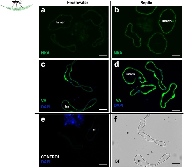
Figure 10.
Na+-K+-ATPase (NKA) and V-type H+-ATPase (VA) immunolocalization in the anal papillae (AP) of wild-collected A. aegypti larvae reared in freshwater (FW) and septic water (Septic). NKA immunostaining (green) of representative transverse sections of the AP from (a) FW-reared larvae and (b) Septic-reared larvae. VA immunostaining (green) of representative transverse sections of the AP from (c) FW-reared larvae and (d) Septic-reared larvae. (e) Control sections of AP (CONTROL, primary antibody omitted). DAPI staining of nuclei is in blue. (f) Representative bright field (BF) image of AP transverse sections in C. Scale bars: 50 µm. Lumen (lm), anal papillae (AP).

