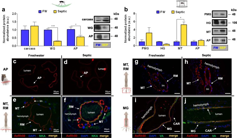
Figure 9.
Rh protein (AeRh50) abundance and immunolocalization in the alimentary canal, anal papillae, and carcass of wild-collected and laboratory A. aegypti larvae reared in freshwater (FW) and septic water (Septic). (a) AeRh50 abundance and representative Western blots (right panel) in the epidermis, whole gut (WG), and anal papillae (AP) of wild A. aegypti larvae (n = 3). (b) AeRh50 abundance and representative Western blots (right panel) in the posterior midgut (PMG), hindgut (HG), Malpighian tubules (MT) and anal papillae (AP) of laboratory A. aegypti larvae (n = 3 FW, n = 4 Septic). The abundance of AeRh50 protein was normalized to total protein (Coomassie protein stain, not shown), and Septic values are expressed relative to the control FW group (assigned a value of 1). Data shown as mean ± S.E.M. Asterisks indicate statistical significance (*p < 0.05; ***p < 0.001) compared to FW control (Unpaired, two-tailed t-test). Representative transverse and cross sections of the anal papillae (AP), Malpighian tubules (MT) and rectum (RM) showing AeRh50 (red) immunostaining from (c–e) wild FW-reared larvae, (d–f) wild Septic-reared larvae. Representative cross sections of the posterior midgut (MG), Malpighian tubules, rectum (RM), and carcass (CAR) showing AeRh50 (red) immunostaining from (g–i) laboratory FW-reared larvae and (h–j) laboratory Septic-reared larvae. Nuclei are labelled by DAPI (blue) staining. Immunostaining of Na+-K+-ATPase (NKA) (e,f) and the V1 subunit of V-type H+-ATPase (VA) (i–j) are shown in green. Co-localization of AeRh50 with V1 subunit of V-type H+-ATPase is indicated (dashed arrows) (merge, yellow). Control sections (primary antibodies omitted, not shown) were devoid of red and green staining. Illustrations of the alimentary canal and anal papillae of A. aegypti larvae to the left of each immunofluorescence image indicates the region of the cross or transverse section (red rectangles). Lumen (lm), midgut (MG), Malpighian tubule (MT), anal papillae (AP); rectum (RM). Scale bars: 100 µm, unless specified.

