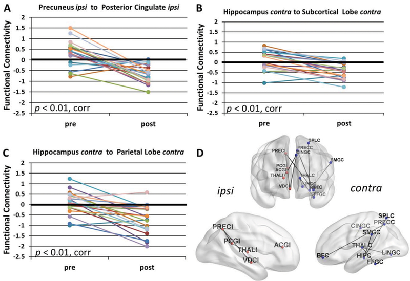
FIG. 2.
FC decreases after surgery in mTLE. Postsurgical decreases in FC compared to presurgical FC are found (A) from the ipsilateral precuneus to the ipsilateral posterior cingulate (p < 0.01, Bonferroni correction); (B) from the contralateral hippocampus to the contralateral subcortical lobe (p < 0.01, Bonferroni correction); and (C) from the contralateral hippocampus to the contralateral parietal lobe (p < 0.01, Bonferroni correction). D: All regional connections with a postsurgical decrease in FC compared to presurgical FC (p < 0.05, Bonferroni correction) are depicted on the brain to indicate the spatial distribution of the changes, excluding those involving the ipsilateral temporal lobe. All statistics are adjusted for months after surgery at which postsurgical scan was obtained. Value of FC = 0 represents age-matched healthy control. ipsi, I (following abbreviation) = ipsilateral to seizure focus; contra, C (following abbreviation) = contralateral to seizure focus; corr = Bonferroni correction; ACG = anterior cingulate gyrus; BF = basal forebrain; CING = mid-cingulate gyrus; FFG = fusiform gyrus; HIP = hippocampus; LING = lingual gyrus; PCG = posterior cingulate gyrus; PREC = precuneus; SMG = supramarginal gyrus; SPL = superior parietal lobule; THAL = thalamus; VDC = ventral diencephalon.

