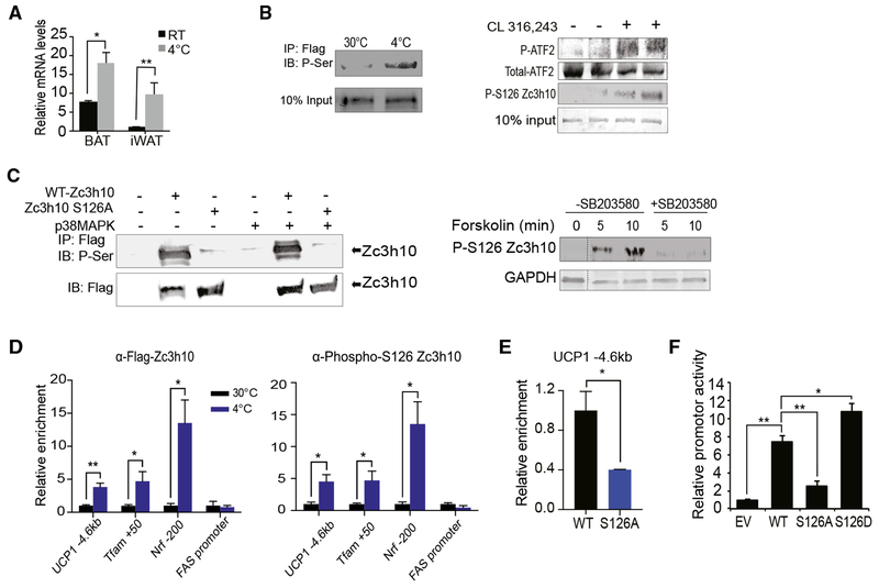
Figure 5. Phosphorylation of Zc3h10-S126 by p38 MAPK for UCP1 Transcription during Cold Exposure.
(A) qRT-PCR for Zc3h10 in BAT/iWAT 10-week-old C57BL76 mice housed at RT or at 4°C (n = 4).
(B) (Left) Immunoblotting of BAT from AQ-Zc3h10 TG mice housed at indicated ambient temperatures using a P-Serine antibody. (Right) Immunoblotting of BAT from AQ-Zc3h10 TG mice that are injected with saline or CL 316,243 using a P-S126 Zc3h10 specific antibody. p38MAPK activity was detected by P-ATF2.
(C) (Left) Immublotting for P-Serine of lysates of HEK293FT cells. (Right) HEK293FT cells were treated with p38 MAPK inhibitor SB203580 (10 μM) for 30 min before the forskolin (1 μM) treatment for the indicated duration.
(D) ChIP-qPCR for Zc3h10 and P-S126 Zc3h10 occupancy to Zc3h10 target genes using BAT chromatin from AQ-Zc3h10 TG.
(E) ChIP-qPCR for Zc3h10 binding at the −4.6 kb region of UCP1 promoter using FLAG antibody.
(F) UCP1 promoter-Luc activity using HEK293FT cells (n = 4).
Data presented as mean ± SEM. *p < 0.05, **p < 0.01, ***p < 0.001. See also Figure S4.

