Fig. 1.
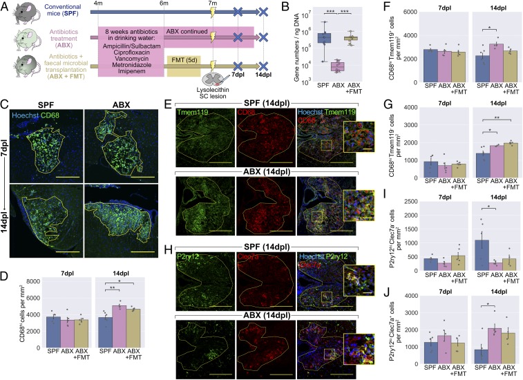
Antibiotics treatment to deplete the microbiota alters the inflammatory response following lysolecithin-mediated demyelination. (A) Mice were administered ABX in their drinking water for 2 mo, after which one group received an FMT while another group were continued on ABX. Mice were killed at 7 and 14 d following lysolecithin injection into the ventral white matter of the spinal cord. (B) RT-PCR of fecal DNA showing depletion of the microbiota by ABX and return to normal levels with FMT. (C) Representative images and (D) density of CD68hi activated microglia/macrophages within the lesion boundary (yellow line). (E) Representative images and density of (F) Tmem119+CD68hi microglia-derived and (G) Tmem119−CD68hi monocyte-derived CD68hi cells within lesions. (H) Representative images and density of (I) P2ry12hiClec7a− homeostatic and (J) P2ry12loClec7a+ degeneration-associated microglia/macrophages within lesions. Insets in E and H are a 3× magnification of the boxed regions. (Scale bars: C, 250 µm; E and H, 200 µm, and Insets, 25 µm.) Error bars show mean ± SEM; *P < 0.05, **P < 0.01, ***P < 0.001; in B, Kruskal−Wallis with Dunn’s post hoc test; in D, F, G, I, and J, 1-way ANOVA with Tukey HSD post hoc test, n = 4 to 6 mice.

