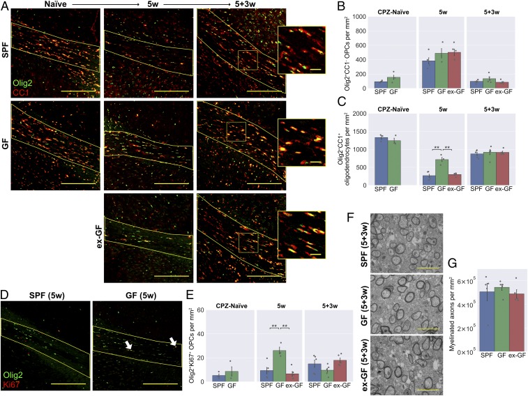
Fig. 5.
GF mice have reduced oligodendrocyte loss following cuprizone administration, but no difference in OPC differentiation. (A) Representative images and density of (B) Olig2+CC1− OPCs and (C) Olig2+CC1+ mature oligodendrocytes within the corpus callosum in cuprizone-naïve mice, following 5 wk cuprizone exposure (5w) and following a further 3 wk of normal diet (5+3w). Insets in A are a 2.5× magnification of the boxed regions. (D) Representative images and (E) density of Olig2+Ki67+ proliferating OPCs within the corpus callosum. Arrow heads in D show representative double-positive cells. (F) Representative electron microscopy images and (G) quantification of myelinated axons within the corpus callosum 3 wk after cuprizone cessation. (Scale bars: A, 200 μm, and Insets, 25 μm; D, 200 μm; F, 2 μm.) Error bars show mean ± SEM; **P < 0.01; 1-way ANOVA, n = 3 to 5 mice.

