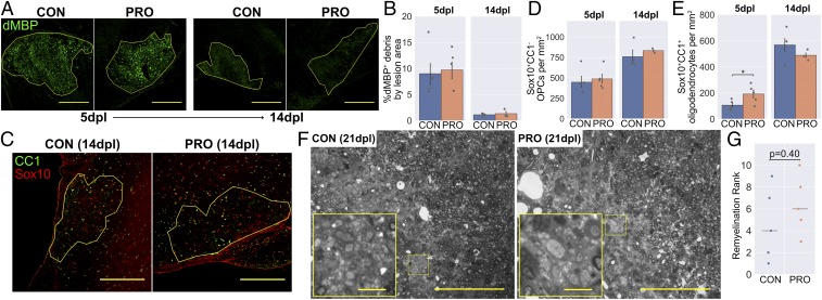
Fig. 7.
VSL#3 probiotic does not enhance remyelination in aged mice. (A) Representative images and (B) area of dMBP+ myelin debris within lesions. (C) Representative images and density of (D) Sox10+CC1− OPCs and (E) Sox10+CC1+ mature oligodendrocytes within lesions. (F) Representative images of toluidine blue-stained resin sections demonstrating persistent demyelination, typical of aged mice (control [CON] rank: 4/10, probiotic [PRO] rank: 3/10). Images are shown in grayscale. Insets are a 4× magnification of the boxed regions. (G) Remyelination ranks assigned by a blinded assessor, with horizontal lines showing the median for each group. (Scale bars: A and C, 250 μm; F, 100 μm, and Insets, 10 μm.) Error bars show mean ± SEM; *P < 0.05; in B, D, and E, Student’s t test, n = 3 to 5 mice; in G, Mann–Whitney U test, n = 5 mice.

