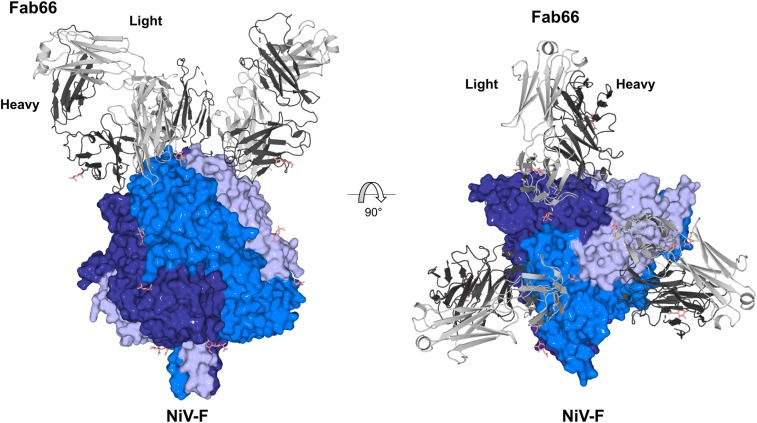
Fig. 1.
Crystal structure of the NiV-F−Fab66 complex. Side and top views show Fab66 bound to an epitope near the apex of the prefusion, uncleaved NiV-F trimer. Each Fab66 molecule binds to a single F protomer in the trimer. The NiV-F trimer is shown as surface with each protomer in the trimer colored a different shade of blue. Fab66 is shown as cartoon and the light and heavy chains are shown in light and dark gray, respectively. N-linked glycans are depicted as sticks and colored salmon.

