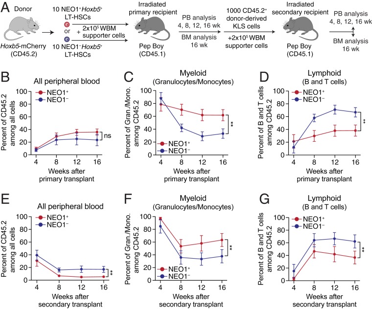
Fig. 4.
NEO1+Hoxb5+ LT-HSCs exhibit myeloid bias and reduced reconstitution potential upon serial transplantation. (A) Experimental design for primary and secondary transplantations of NEO1+ and NEO1− Hoxb5+ LT-HSCs. BM, bone marrow. (B–D) Measurements of reconstitution potential and lineage priming after primary transplantation (“NEO1+,” n = 14 mice; “NEO1−,” n = 11 mice) from 2 independent experiments. (B) Percent of donor-derived (CD45.2+) cells among all peripheral blood cells at 4, 8, 12, and 16 wk posttransplant. (C) Percent of myeloid cells (GR1+CD11B+ granulocytes and monocytes) among donor-derived (CD45.2+) cells at 4, 8, 12, and 16 wk posttransplant. (D) Percent of lymphoid cells (B220+ B cells and CD3+ T cells) among donor-derived (CD45.2+) cells at 4, 8, 12, and 16 wk posttransplant. (E–G) Same as in B–D but analyzing peripheral blood in secondary recipients transplanted with 1,000 donor-derived Lin−c-KIT+SCA1+ (KLS) cells from primary hosts (“NEO1+,” n = 8; “NEO1−,” n = 9) from 2 independent experiments. Statistical significance for B–G was calculated using 2-way ANOVA with time posttransplant and NEO1 status as factors. **P < 0.01; ns, nonsignificant. All line plots in this figure indicate mean ± SEM.

