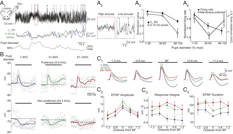
Fig. 3.
Elevated arousal reduces membrane potential variability, spontaneous firing, and lateral inhibition. (A1, Top) Current-clamp recording of membrane potential (Vm) in a representative L2/3 cell. Asterisks mark truncated action potentials. (Middle) Vm SD (purple) and 2–10 Hz power (green) over 1 s intervals. (Bottom) Pupil diameter. (A2) Expansion of areas marked in A1. (A3) Summary showing that as arousal increases, 2–10 Hz power (open circles) and SD (filled circles) decrease. (A4) Summary showing that spontaneous firing decreases as arousal increases (filled circles, n = 19). Mean Vm is most hyperpolarized during moderate arousal (open circles, n = 30 cells). (B) Responses to a preferred (Top) and nonpreferred (Bottom) tone (black bar) during different arousal levels in a representative cell. Gray, subset of single trials. Bold, mean response. Dashed line, baseline Vm. (C1) Average responses to tones aligned to BF of each cell during low (blue), moderate (green), and high (red) arousals (n = 15 cells). Dashed line, baseline Vm. (C2) Arousal causes a modest increase in EPSP peak amplitude. (C3) Responses shift from net hyperpolarization to net depolarization for tones ≥BF. (C4) Arousal-dependent suppression of lateral inhibition increases EPSP duration. Error bars, SEM.

