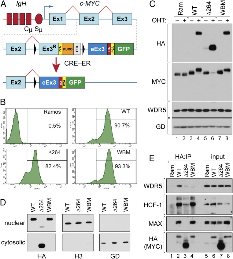
Fig. 1.
Inducible c-MYC exon swap in a BL cell line. (A) Structure of the c-MYC gene in Ramos cells (Top). Chromosome 14 is in red and shows part of the IgH locus including the constant (Cµ) and switch (Sµ) regions. Chromosome 8 is depicted in blue and shows exons 1 to 3 of c-MYC (Ex1–Ex3). Schematic of the modified locus in the unswitched state (Middle). Ex3R; recoded WT exon 3. P2A, “self-cleaving” peptide; PURO, puromycin resistance gene; TER, transcriptional terminator; eEx3, exchanged exon 3 (encoding WT, WBM, or ∆264 MYC sequences); HA, hemagglutinin epitope tag. LoxP sites are the black triangles. Configuration of the altered locus after activation of ER-linked CRE recombinase (Bottom). (B) Cells were treated with 20 nM OHT for 24 h, and GFP-positive cells scored by flow cytometry. The percentage of GFP-positive cells in each population is shown. (C) Western blot, performed on lysates from parental Ramos CRE–ER expressing cells (Ram), or modified cells expressing the WT, WBM, or ∆264 c-MYC proteins. Cells were untreated (−) or treated (+) with 20 nM OHT for 24 h prior to lysate preparation. Blots were probed with anti-HA, anti-MYC, anti-WDR5, and anti-GAPDH (GD) antibodies as indicated. (D) Engineered Ramos cells were switched to express the indicated MYC protein and subject to biochemical fractionation into nuclear and cytosolic extracts. Extracts were then probed with antibodies against HA (switched MYC), histone H3 (H3; nuclear), or GAPDH (GD; cytosolic). (E) Cell lines were treated with 20 nM OHT (24 h), lysates prepared, and HA-tagged MYC proteins immunoprecipitated (IP) by an anti-HA antibody. Immune complexes (lanes 1 through 4) were then probed for WDR5, HCF-1, MAX, or HA-tagged MYC by Western blotting. A sample of the lysates (input) were also probed by Western blot (lanes 5 through 8). This IP is representative of 3 independent experiments.

