Fig. 4.
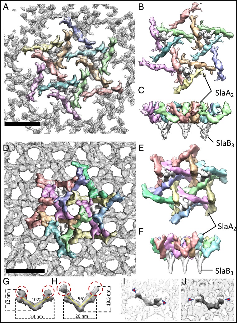
Assembly models for the S. acidocaldarius (A–C) and S. solfataricus (D–F) S-layers. (A and D) Outward facing surfaces (gray mesh) segmented into SlaA dimers (each dimer in a different color) and SlaB trimers (white). (B and E) Inward facing surface. (C and F) Side view, perpendicular to the membrane plane. (G–J) Comparison of the structures of SlaA from Saci (G) and Ssol (H) show differences in length, height, and angular shape of the dimer. (I and J) Location of 1 SlaA dimer within the S-layer of Saci (I) and Ssol (J). Red circles/arrowheads indicate apical domains that determine shape and topology of the hexameric pores. (Scale bars, 20 nm).

