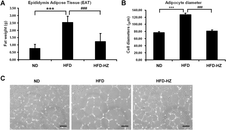
Fig. 4.
The effect of HZ extract treatment on epididymis adipose tissue (EAT) in C57BL/6 J mice fed an HFD. a The weight of EAT. b The diameters of adipocytes. c hematoxylin-eosin staining of adipocytes in the EAT of mice. The scale bar is 100 μM. Data are shown as means ± SEM (n = 10 per group). ND vs. HFD: *p < 0.05; **p < 0.01; ***p < 0.001. HFD vs. HZ: #p < 0.05; ##p < 0.01; ###p < 0.001

