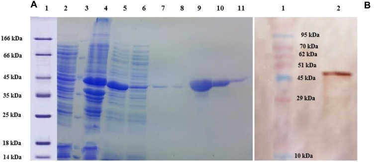
Figure 1.
(A) SDS PAGE (12%) gel showing expression and purification of lysostaphin. Lane 1: molecular weight marker, Lane 2: uninduced cell extract, Lane 3: molecular weight marker, Lane 4: proteins from pellet after separation of readily soluble protein fraction, Lane 5: readily soluble protein extract (cytoplasmic cell proteins), Lane 6: flow through Ni-NTA agarose resin affinity chromatography, Lanes 7 and 8: Non-tagged proteins washed from affinity chromatography by 20 mM imidazole, Lanes 9–11: lysostaphin eluted by 250 mM imidazole. (B) Western Blotting analysis using anti-His-HRP conjugated antibody. Lane 1: molecular weight marker, Lane 2: lysostaphin.

