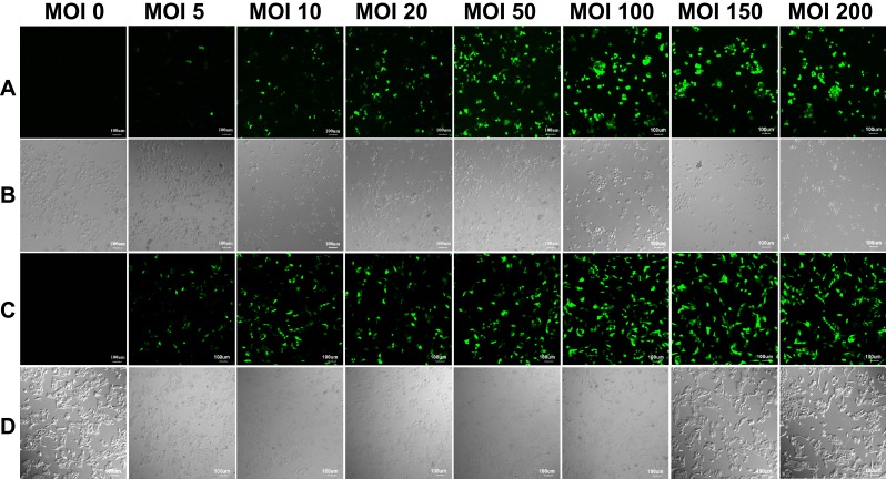
Figure 3.
The laser confocal fluorescence images of SMMC-7721 and L-02 cells infected with Ad at MOIs of 0, 5, 10, 20, 50, 100, 150 and 200, respectively. (A) Fluorescence field and (B) bright-field images of SMMC-7721 cells; (C) fluorescence field and (D) bright-field images of L-02 cells. Scale bar: 100 μm.
Abbreviations: Ad, adenovirus; MOI, multiplicity of infection.

