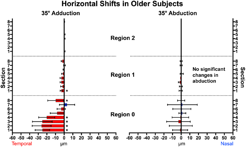
Fig. 6.
Horizontal displacement in older subjects during adduction (left) and abduction (right). In adduction, the nasal half (Sections 1–4) of the optic nerve head (Region 0) exhibited greatest temporal shift while there were smaller temporal displacements in Region 1. There were no significant displacements in abduction. * signifies non-zero displacements. Brackets mark 95% confidence intervals.

