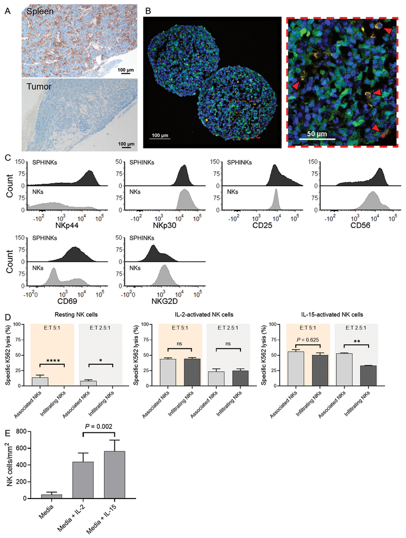
Figure 3.
Phenotype and function of sphere-infiltrating NK cells (SPHINKs). A, In vivo infiltration of spleen by adoptively transferred IL-15–activated NK cells in NOD-SCID IL2Rγnull mice. Immunohistochemistry staining for human CD45 is shown. In the same animal, IL-15–activated NK cells did not infiltrate the tumor microenvironment (SJNBL046_X) B, YFP-expressing PDX cells (green) were used to form tumorspheres. IL-15–activated NK cells (orange) are capable of infiltrating tumorspheres (red arrows) as early as 4 hours after the assay start. DAPI staining was performed for nuclear discrimination (blue). Experiments were performed with two donors that yielded comparable findings. Representative results from one donor are shown. C, Phenotypic characterization of IL-15–activated SPHINKs and IL-15–activated noninfiltrating (i.e., tumor-associated) NK cells by flow cytometry. D, Resting tumorsphere-infiltrating NK cells exhibited decreased cytotoxicity against K562 than did tumorsphere-non-infiltrating NK cells. This was also noted for IL-15-activated NK cells at an E:T ratio of 2.5:1. At an E:T ratio of 5:1 and when NK cells were pre-activated with IL-2, preserved cytotoxicity was noted. The experiments were performed in technical triplicates with pooled SPHINKs from 30 tumorspheres. The mean specific lysis and standard deviation were plotted. E, The number of SPHINKs was higher when NK cells were activated with IL-15 than with IL-2 or media alone.

