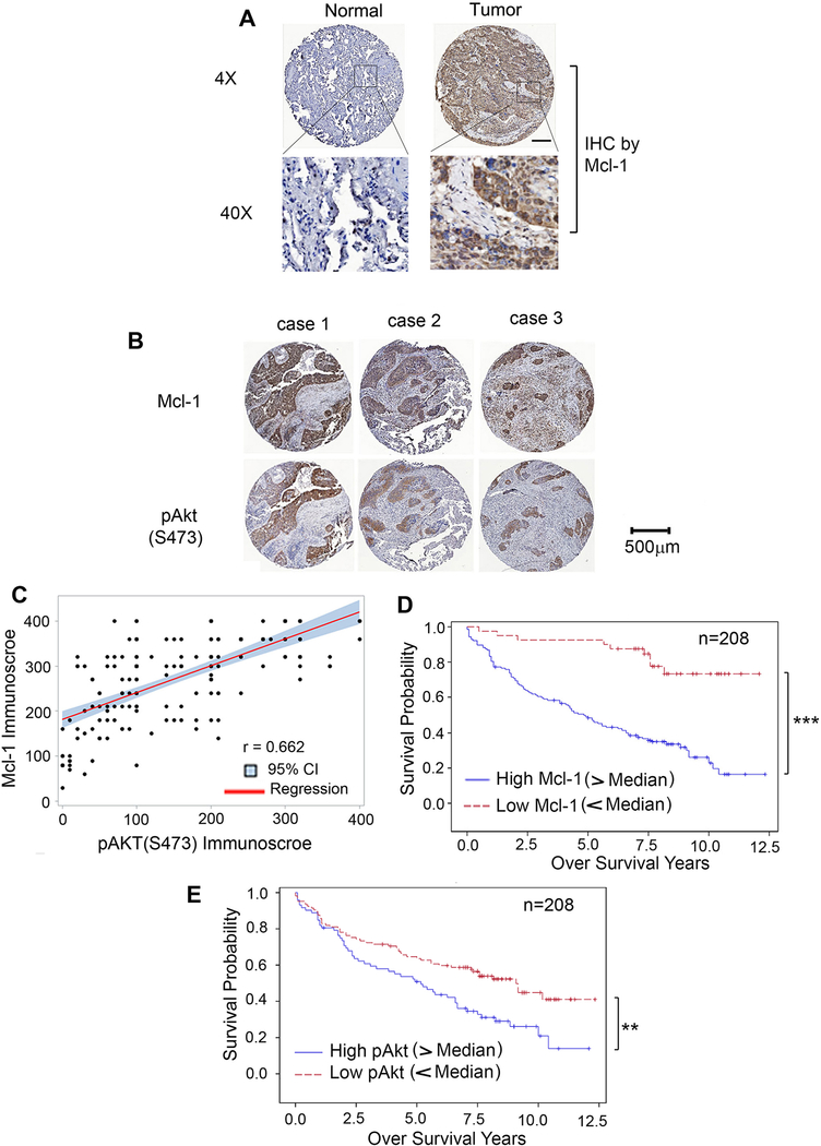
Figure 5.
Mcl-1 and pAkt are potential prognostic biomarkers for patients with NSCLC. A, Mcl-1 expression in normal lung tissues versus lung cancer tissues from a representative NSCLC case was analyzed by IHC using Mcl-1 antibody. Normal tissue is the adjacent normal lung tissue from the same case. Mcl-1 expression was quantified by immunoscore. B, IHC staining of Mcl-1 and pAkt (S473) was compared in tumor tissues from 3 representative NSCLC cases. C, The correlation between Mcl-1 and pAkt immunoscores in NSCLC patient tumors (n = 208) was explored using Pearson correlation analysis. Scatter plots of Mcl-1 immunoscore and pAkt immunoscore were produced showing the fitted linear regression lines and 95% confidence intervals. D and E, Kaplan-Meier survival curves of NSCLC patients: low Mcl-1 vs. high Mcl-1 (D), low pAkt vs. high pAkt (E), n = 208. ***P < 0.001, **P < 0.01, by log-rank test. Low: immunoscore < 200, high: immunoscore > 200.

