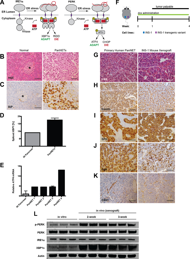
Figure 1. PanNETs show evidence of ER stress and UPR activation.
A, In response to the accumulation of misfolded proteins in the ER, IRE1α and PERK homodimerize and signal an adaptive stress response through splicing of Xbp1 and phosphorylation of eIF2α, respectively. However, under sustained ER stress, these pathways promote apoptosis through RIDD and upregulation of pro-apoptotic CHOP.
B-C, Representative (B) H&E and (C) BiP/GRP78 IHC on normal pancreas and primary human PanNET. Star indicates islet of Langerhans (scale bars, 50 μm).
D-E, (D) Percent XBP1 splicing and (E) relative ATF4 mRNA expression from normal human pancreas and four primary human PanNETs. Technical replicate error bars shown in E.
F, PanNET xenograft experimental setup. INS-1 cells (control vs. transgenic variant) injected s.c. in bilateral flanks of NSG mice. Tumors become palpable by ~10 d; mice are sacrificed at 4 weeks post-injection.
G-K, IHC of human PanNETs and INS-1 mouse xenografts stained with the indicated antibodies (CgA=chromogranin A, SPH=synaptophysin; scale bars, 50 μm).
L, INS-1 cells were grown in tissue culture (in vitro) or as xenografts (in vivo) in NSG mice for 2 or 3 weeks. Three unique replicates of each condition were harvested and analyzed by immunoblotting with the indicated antibodies. Quantified in Fig. S1B and C.

