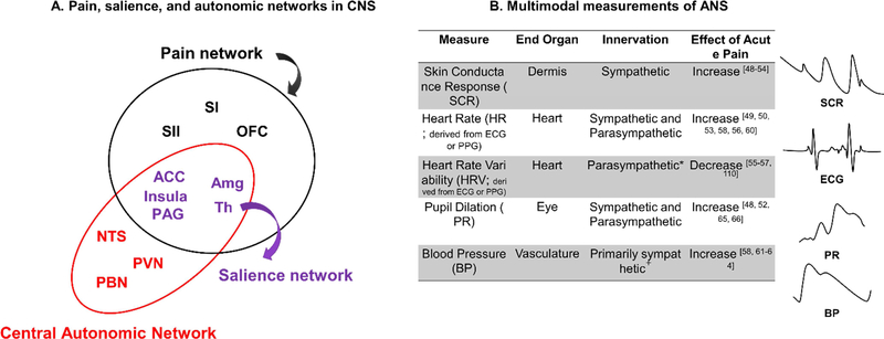
Figure 2. Measurements of brain and bodily responses to pain.
A. Anatomical overlap between pain (black and purple regions), salience (purple regions), and autonomic networks (red and purple regions) in central nervous system. Amg: amygdala; ACC: anterior cingulate cortex; NTS: nucleus tractus solitarius of the medulla; OFC: orbitofrontal cortex; PAG: periacqueductal gray; PBN: parabrachial nuclei; PVN: paraventricular nucleus of the hypothalamus; SI: primary somatosensory cortex; SII: secondary somatosensory cortex; Th: thalamus.
B. Various ANS measurements (e.g., skin conductance response, heart rate, heart rate variability, pupil dilation, blood pressure) across different end-organs are related to pain. Because these end-organs are innervated by different branches of the autonomic nervous system and influenced by different mechanisms in the periphery, they provide non-overlapping information about arousal, and are best used in combination. *Many measures of HRV are parasympathetically mediated, but some measures are jointly influenced by both the sympathetic and parasympathetic nervous system. +Blood pressure in the vasculature is primarily controlled by the sympathetic nervous system, but parasympathetic control of the heart also influences blood pressure.

