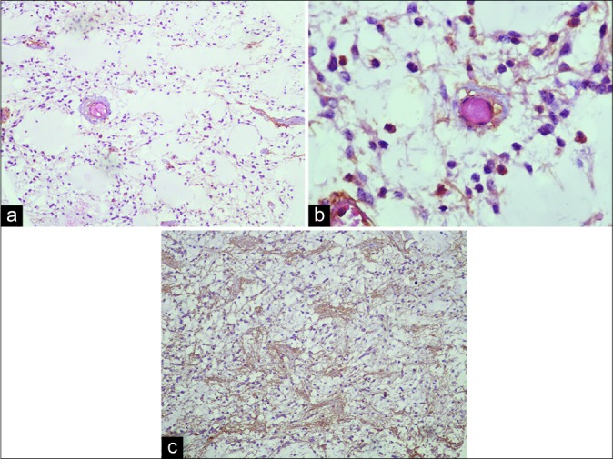
Figure 2:

This shows histopathology of resected tumor. We could see hypocellular glial tumor, in which monomorphic piloid tumor cells are dispersed in a myxoid matrix (Hematoxylin and Eosin stain, ×100) (a). Furthermore, tendency of piloid perivascular cells to form a rosette-like structure without any Rosenthal fiber could be seen (Hematoxylin and Eosin stain, ×400, b). (c) Reveals a strong immunoreactivity of the tumor cells for glial fibrillary acidic protein (GFAP immunostaining, ×100, c).
