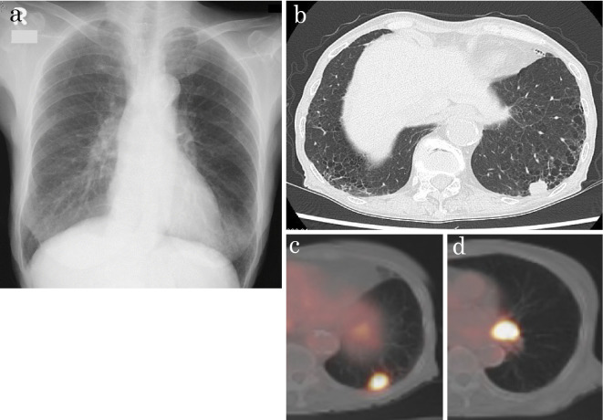
Figure 1.
(a) Chest X-ray showing a mass shadow at the left lower lung field. (b) Chest CT scan showing a nodule with notching and spicula formation under the pleural cavity in the left lower lobe and lymphadenopathy in the left hilum of the lung. (c) Whole-body positron emission tomography showing the lung lesion, with an SUVmax of 11.6 for the nodule in the left lower lobe. (d) Whole-body positron emission tomography showing the lymph node with an SUVmax of 18.4 in the left hilum of the lung. CT: computed tomography, SUV: standardized uptake value

