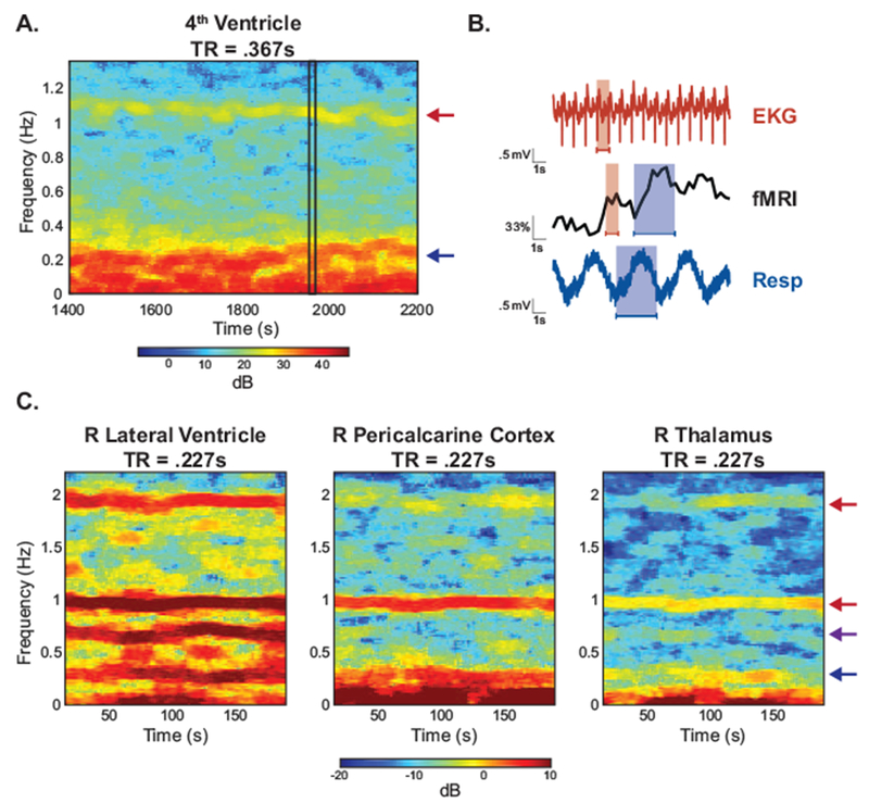
Figure 1: Physiological noise sampled directly in fast fMRI.

Unlike conventional fMRI, physiological noise can be resolved without aliasing in fast fMRI. (A) A spectrogram of the 4th ventricle from a subject in Experiment A shows high power oscillations in the cardiac (red arrow) and respiratory (blue arrow) frequency range. (B) A zoomed-in time series from (A) (black rectangle) shows that the high-power oscillations correspond to cardiac (red) and respiratory (blue) cycles obtained from external physiological recordings. (C) Spectrograms from ROIs in Experiment B manifest the harmonic structure of the physiological noise. In the right lateral ventricle (left) one respiration term (blue arrow), two cardiac terms (red arrow), and one interaction term (purple arrow) are observed. These components are also present to varying degrees in pericalcarine cortex (middle) and the thalamus (right).
