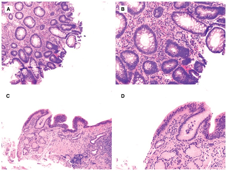
Figure 3.
Low-grade dysplasia in inflammatory bowel disease (IBD)- and Barrett’s esophagus (BE)-surveillance biopsies. (A) and (B) IBD colonic biopsy shows focal area resembling tubular adenoma, consisting of glands lined by enlarged, stratified, hyperchromatic nuclei (H&E stain; (A) magnification 100×; (B) magnification 200×). The changes extend to the surface epithelium (lack of surface maturation). There is no architectural complexity, loss of nuclear polarity, or significant pleomorphism—features to suggest HGD. (C) and (D) Similar features are present in a BE-surveillance biopsy, supporting the diagnosis of low-grade dysplasia (H&E stain; (C) magnification 40×; (D) magnification 100×).

