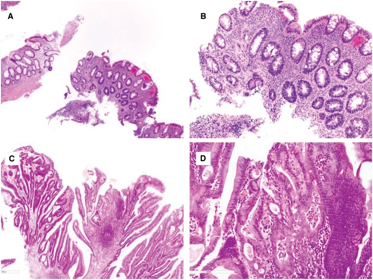
Figure 4.
Chronic colitis with high-grade dysplasia. (A) and (B) This biopsy shows an area consisting of atypical glands lined by enlarged dark nuclei without surface maturation (H&E stain; (A) magnification 100×; (B) magnification 200×). The adjacent fragment provides an internal negative control. (C) and (D) This colon biopsy shows architectural and cytologic features of high-grade dysplasia (H&E stain; (C) magnification 40×; (D) magnification 200×). There is focal cribriforming, nuclear pleomorphism, and loss of nuclear polarity.

