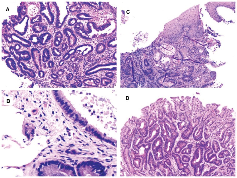
Figure 8.
Barrett’s esophagus with features suspicious for carcinoma. (A) This biopsy shows features of high-grade dysplasia (HGD). In addition, there are a few glands with eosinophilic luminal debris (H&E stain, magnification 100×). (B) This biopsy shows HGD. In addition, there is one single cell infiltrating into the lamina propria (H&E stain, magnification 100×). (C) This biopsy shows HGD. In addition, there are a few glands with eosinophilic luminal debris and incorporation of neoplastic glands onto the squamous epithelium (H&E stain, magnification 200×). (D) This biopsy shows HGD. In addition, there is back-to-back glandular crowding (H&E stain, magnification 100×). All these features are worrisome for, but not diagnostic of, intramucosal adenocarcinoma.

