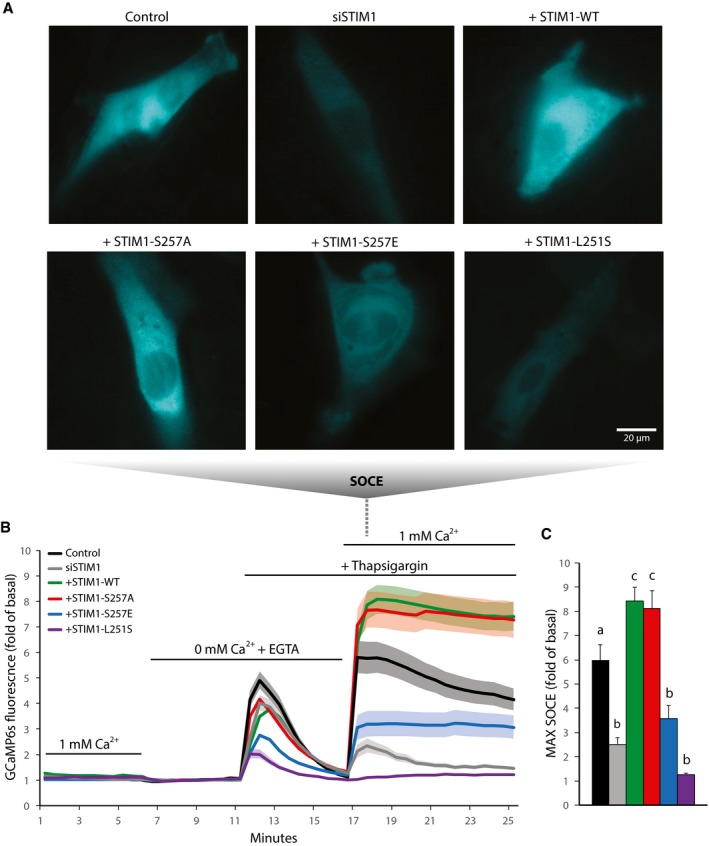
Figure 5. Phosphorylation of STIM1 by AMPK suppresses SOCE .

-
A–CL6 myoblasts were transfected with the genetic cytosolic Ca2+ sensor GCaMP6s. Cells were also transfected with non‐targeted siRNA (Control) or siRNA directed at endogenous STIM1 (siSTIM1). Some cells with endogenous STIM1 knockdown were also transfected with full‐length wild‐type (WT) or mutant (S257A, S257E, or L251S) STIM1‐mRuby3. (A) Representative microscopy images of GCaMP6s signal indicating SOCE in L6 myoblasts, (B) Ca2+ levels over time (fold of 0 mM Ca2+ + EGTA basal), and (C) quantification of SOCE in L6 myoblasts incubated in buffer containing 1 or 0 mM Ca2+ and treated with 2 μM thapsigargin during the indicated times. For (C), “b” is significantly different from “a” and “c”, and “c” is significantly different from “a” and “b”, P < 0.05 by one‐way ANOVA with Tukey's post‐hoc test. For (B‐C), data are represented as mean ± SEM, n = 5 independent experiments (n = 3 for L251S).
Source data are available online for this figure.
