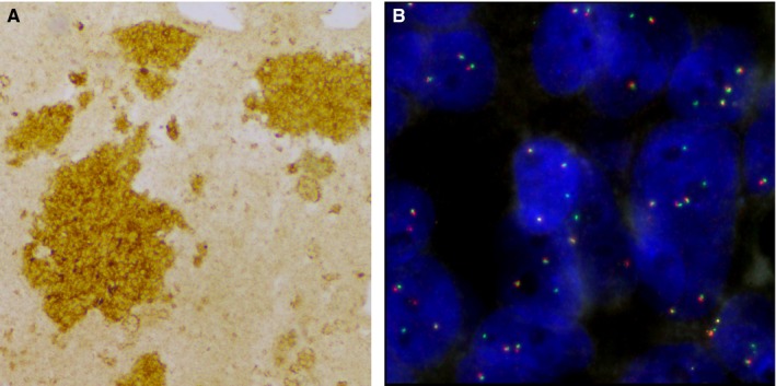
Figure 1.

IHC and FISH analyses of ALK rearrangement of E5844 tumor. A, Photograph of IHC cytoplasmic staining in more than 10% of tumor cells at a score of 1+ (×10). B, Photograph of FISH showing atypical pattern of fused signal associated with isolated green signal (×100). FISH, fluorescence in situ hybridization; IHC, immunohistochemistry
